 |
|||||
 |
 |
||||
 |
|||||
 |
 |
||||

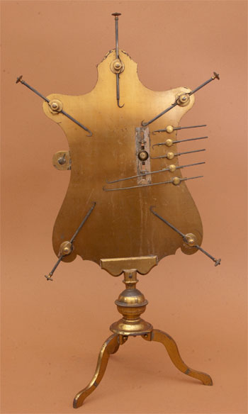
Johan Georg Mitsdörffer’s large dissecting microscope was based on a form developed by Berlin physician Johann Lieberkühn in the first half of the eighteenth century. Lieberkühn dubbed this instrument an “anatomical microscope,” but it is better described as a dissecting microscope. It was used for studying the membranes and circulation of blood in small animals. The microscope employed a thin sheet of brass formed in the shape of an animal figure, with ten sliding hooks positioned around its perimeter. Four larger hooks at the corners were designed to hold the legs of the live subject, while the smaller hooks were used to pull back successive layers of membrane opposite the lens for examining its tissue and circulation. To achieve focus, a thumbscrew adjusted the pliable band to which the lens was secured. This microscope required strategic placement before a window or lamp for light to pass through the examined material. The entire instrument is attached to a ball-and-socket joint and is supported on tripod legs. The instrument can be stored in a mahogany case. The microscope is 52cm tall.
This instrument is described in the 1768 three volume set by Martin Frobéne Ledermuller entitled "Troisiéme Cinquantaine des Amusemens Microscopiques". There is one other known instrument in the Science Museum of London.
(Thanks to Timothy O'Brien of the SFO Museum for this description).
Featured 01/2012